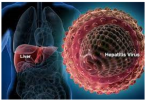
VIRAL HEPATITIS
The term viral hepatitis is used to describe infection of the liver caused by hepatotropic viruses. Currently there are 5 main varieties of these viruses and a sixth poorly characterised virus, causing distinct types of viral hepatitis:
Hepatitis A virus (HAV), causing a faecally-spread self-limiting disease;
Hepatitis B virus (HBV), causing a parenterally transmitted disease that may become chronic;
Hepatitis C virus (HCV), previously termed non-A, non-B (NANB) hepatitis virus involved chiefly in transfusion-related hepatitis;
Hepatitis delta virus (HDV) which is sometimes associated as superinfection with hepatitis B infection;
Hepatitis E virus (HEV), causing water-borne infection; and
Hepatitis G virus (HGV), is a recently discovered parenterally transmitted hepatotropic virus.
All these human hepatitis viruses are RNA viruses except HBV which is a DNA virus.
Though a number of other viral diseases such as infection with Epstein-Barr virus (in infectious mono nucleosis), arbovirus (in yellow fever), cytomegalovirus, herpes simplex and several others affect the liver but the changes produced by them are nonspecific and the term ‘viral hepatitis’ is strictly applied to infection of the liver by the hepatitis viruses.
ETIOLOGIC CLASSIFICATION
Based on the etiologic agent, viral hepatitis is currently classified into 6 etiologic types-hepatitis A, hepatitis B, hepatitis C, hepatitis D, hepatitis E and hepatitis G.
Hepatitis A
Infection with HAV causes hepatitis A (infectious hepatitis). Hepatitis A is responsible for 20-25% of clinical hepatitis in the developing countries of the world but the incidence is much lower in the developed countries. Hepatitis A is usually a benign, self-limiting disease and has an incubation period of 15-45 days. The disease occurs in epidemic form as well as sporadi cally. It is usually spread by faeco-oral route. Parenteral transmission is extremely rare. The spread is related to close personal contact such as in overcrowding, poor hygiene and poor sanitation. Most frequently affected age is 5-14 years; adults are often infected by spread from children.
HEPATITIS A VIRUS (HAV).
The etiologic agent for hepatitis A, HAV, is a small, 27 nm diameter, icosahedral non-enveloped, single-stranded RNA virus. Viral genome has been characterised but only a single serotype has been identified. HAV infection can be transmitted to primates and the virus can be cultivated in vitro. Inactivation of viral activity can be achieved by boiling for 1 minute, by ultraviolet radiation or by contact with formaldehyde and chlorine. The virus is present in the liver cells, bile, stool and blood during the incubation period and in pre-icteric phase but viral shedding diminishes after the onset of jaundice. Chronic carriers have not been identified for HAV infection.
PATHOGENESIS.
The mechanism by which HAV infection causes hepatitis A is poorly understood. An immunologic basis is suspected but the evidence in support is indirect in the form of immunologic markers but not direct demonstration of the etiologic agent in the affected hepatocytes. These markers are :
- IgM anti-HAV antibody appears in the serum at the onset of symptoms of acute hepatitis A.
- IgG anti-HAV antibody is detected in the serum after IgM antibody and gives life-long protective immunity against reinfection with HAV.
Hepatitis B
Hepatitis B (serum hepatitis) caused by HBV infection has a longer incubation period (30-180 days) and is transmitted parenterally such as in recipients of blood and blood products, intravenous drug addicts, patients treated by renal dialysis and hospital workers exposed to blood, and by intimate physical contact such as from mother to child and sexually. The disease may occur at any age. HBV infection causes more severe form of illness that includes: acute hepatitis B, chronic hepatitis, progression to cirrhosis, fulminant hepatitis and an asymptomatic carrier stage. HBV plays some role in the development of hepatocellular carcinoma as discussed later.
HEPATITIS B VIRUS (HBV). The etiologic agent for hepatitis B, HBV, is a DNA virus which has been extensively studied. Electron microscopic studies on serum of patients infected with HBV show 3 forms of viral particles of 2 sizes-small (spheres and tubules/ filaments) and large (spheres) as under:
- i) Small particles are most numerous and are in two forms: as 22 nm spheres, and as tubules 22 nm in diameter and 100 nm long. These are antigenically identical to envelope protein of HBV and represent excess of viral envelope protein referred as hepatitis B surface antigen (HBsAg).
- ii) Large particles, 42 nm in diameter, are double-shelled spherical particles, also called as Danc particles. These are about 100 to 1000 times less in number in serum compared to small 22 nm particles and represent intact virion of HBV.
The genomic structure of HBV is quite compact and complex. The HBV DNA consists of 4 overlapping genes which encode for multiple proteins :
- S gene codes for the major surface envelope protein, hepatitis B surface antigen (HBsAg). HBsAg is present on the outer surface of the large spherical particles as well as in small spherical and tubular structures. Pre S1 and pre-S2 regions of genome which are upstream of S gene, code for two other large proteins.
- P gene is the largest and codes for DNA polymerase.
- C gene codes for two nucleocapsid proteins, HBeAg and a core protein termed HBcAg.
- X gene codes for HBxAg having a role in activation and transcription of viral and cellular genes and may actually contribute to carcinogenesis by binding with TP53.
PATHOGENESIS. The evidence linking immuno pathogenetic mechanism with hepatocellular damage is much stronger in HBV infection than with HAV infection. In support of immune pathogenesis is the demons tration of several immunological markers (serologic as well as viral), and molecular and morphologic evidence that hepatocytic damage is initiated by virus-infected CD8+T cytotoxic cells.
Serologic and viral markers. Various immunological markers indicative of presence of HBV infection can be demonstrated in the sera as well as in the hepatocytes of infected individuals. These are as under :
- HBsAg. In 1965, Blumberg and colleagues in Philadelphia found a lipoprotein complex in the serum of a multiple-transfused haemophiliac of Australian aborigine which was subsequently shown by them to be associated with serum hepatitis. This antigen was termed Australia antigen by them (In 1977, Blumberg was awarded the Nobel Prize for his discovery). The term Australia antigen is now used synonymous with hepatitis B surface antigen (HBsAg). HBsAg appears early in the blood after about 6 weeks of infection and its detection is an indicator of active HBV infection. It usually disappears in 3-6 months. Its persistence for more than 6 months implies a carrier state. HBsAg may also be demonstrated in the cell membrane of hepato cytes of carriers and chronic hepatitis patients by Orcein staining (orange positivity) but not in the hepatocytes during acute stage of illness.
- Anti-HBs. Specific antibody to HBsAg in serum called anti-HBs appears late, about 3 months after the onset. Anti-HBs response may be both IgM and IgG type. The prevalence rate of anti-HBs ranges from 10-15%. In these individuals it persists for life providing protection against reinfection with HBV.
- HBeAg. HBeAg derived from core protein is present transiently (3-6 weeks) during an acute attack. Its persistence beyond 10 weeks is indicative of develop ment of chronic liver disease and carrier state.
- Anti-HBe. Antibody to HBeAg called anti-HBe appears after disappearance of HBeAg. Seroconversion from HBeAg to anti-HBe during acute stage of illness is a prognostic sign for resolution of infection.
- HBcAg. HBcAg derived from core protein cannot be detected in the blood. But HBCAg can be demons trated in the nuclei of hepatocytes in carrier state and in chronic hepatitis patients by Orcein staining but not in the liver cells during acute stage.
- Anti-HBc. Antibody to HBCAg called anti-HBc can, however, be detected in the serum of acute hepatitis B patients during pre-icteric stage. Anti-HBc may be IgM or IgG class antibody. IgM anti-HBc persists for 4-6 months and is followed later by IgG anti-HBc. Thus detection of high titre of IgM anti-HBc is indicative of recent acute HBV infection, while elevated level of IgG anti-HBc suggests HBV infection in the remote past.
- HBV-DNA. Detection of HBV-DNA by molecular hybridisation using the Southern blot technique is the most sensitive index of hepatitis B infection. It is present in pre-symptomatic phase and transiently during early acute stage.
Hepatitis D
Infection with delta virus (HDV) in the hepatocyte nuclei of HBsAg-positive patients is termed hepatitis D. HDV is a defective virus for which HBV is the helper. Thus, hepatitis D develops when there is concomitant hepatitis B infection. HDV infection and hepatitis B may be simultaneous (co-infection), or HDV may infect a chronic HBsAg carrier (superinfection) :
With coinfection, acute hepatitis D may range from mild to fulminant hepatitis but fulminant hepatitis is more likely in such simultaneous delta infection. Chronicity rarely develops in coinfection.
With superinfection, (incubation period 30-35 days), chronic HBV infection gets worsened indicated by appearance of severe and fulminant acute attacks, progression of carrier stage to chronic delta hepatitis or acceleration towards cirrhosis. Hepatocellular carcinoma is, however, less common in HBsAg carriers with HDV infection.
HDV infection is worldwide in distribution though the incidence may vary in different countries. Endemic regions are Southern Europe, Middle-East, South India and parts of Africa. The high risk individuals for HDV infection are the same as for HBV infection i.e. intra venous drug abusers, homosexuals, transfusion reci pients, and health care workers.
HEPATITIS DELTA VIRUS (HDV).
The etiologic agent, HDV, is a small single-stranded RNA particle with a diameter of 36 nm. It is double-shelled-the outer shell consists of HBsAg and the inner shell consists of delta antigen provided by a circular RNA strand. It is highly infectious and can induce hepatitis in any HBsAg positive host. HDV replication and proliferation takes place within the nuclei of liver cells. Markers for HDV infection include the following:
- HDV identification in the blood and in the liver cell nuclei.
- HDAg detectable in the blood and on fixed liver tissue specimens.
- Anti-HD antibody in acute hepatitis which is initially IgM type and later replaced by IgG type anti-HD antibody which persists for life to confer immunity against reinfection.
PATHOGENESIS. HDV, unlike HBV, is thought to cause direct cytopathic effect on hepatocytes. However, there are examples of transmission of HDV infection from individuals who themselves have not suffered from any attack of hepatitis, suggesting that it may not be always cytopathic. Thus, the exact mechanism remains unresolved.
Hepatitis C
The diagnosis of a third major category of hepatitis was earlier made after exclusion of infection with other. known hepatitis viruses and was designated non-A, non-B (NANB) hepatitis. However, now this third type has been characterised and is called hepatitis C.
Hepatitis C infection is acquired by blood trans fusions, blood products, haemodialysis, parenteral drug abuse and accidental cuts and needle-pricks in health workers. About 90% of post-transfusion hepatitis is of hepatitis C type. About 1-2% of volunteer blood donors and up to 5% of professional blood donors are carriers of HCV. Hepatitis C has an incubation period of 20-90 days (mean 50 days). Clinically, acute HCV hepatitis is milder than HBV hepatitis but HCV has a higher rate of progression to chronic hepatitis than HBV. Persistence of infection and chronic hepatitis are the key features of HCV. Occurrence of cirrhosis after 5 to 10 years and progression to hepatocellular carcinoma are other conse quences of HCV infection. Currently, HCV is considered more important cause of chronic liver disease worldwide than HBV.
HEPATITIS C VIRUS (HCV). HCV is a single-stranded, enveloped RNA virus, having a diameter of 30-60 nm. HCV genome has about 3000 amino acids. The genomic organisation of HCV shows a 5′ terminal end, C (capsid) region and the envelope regions E1 and E2 in the exons
- Anti-HCV antibodies. Three generations of anti-HCV IgG assays are available:
- i) First generation antibodies are against C100-3 region proteins and appear 1 to 3 months after infection.
- ii) Second generation antibodies are against C200 and C33c proteins and appear about one month earlier than the first generation.
iii) Third generation antibodies are against C22-3 and NS-5 region proteins and are detected even earlier.
- HCV-RNA. HCV infection is, however, confirmed by HCV-RNA employing, PCR technique which can be detected within a few days after exposure to HCV infec tion, much before appearance of anti-HCV and persists for the duration of HCV infection.
PATHOGENESIS. How HCV causes liver cell injury is not yet clearly established. HCV virions have not been identified in hepatocytes. Cell-mediated immune mecha nism certainly play a role in hepatocytic injury due to HCV. Perhaps, HCV infection of lymphoid cells may play a role in immunologic injury to hepatocytes. In patients with chronic HCV hepatitis, HCV-specific CD4+ T cells and HLA-restricted CD8+ T cells have been identified. Viral and host crossreacting antibodies to liver and kidney microsomal antigen (anti-LKM) have been reported in a subset of patients that explains the association of autoimmune hepatitis and HCV hepatitis.
Hepatitis E
Hepatitis E is an enterically-transmitted virus, pre viously labelled as epidemic or enterically transmitted type of non-A non-B hepatitis. The infection occurs in young or middle-aged individuals, primarily in India, other Asian countries, Africa and central America.
The infection is generally acquired by contamination of water supplies such as after monsoon flooding. However, compared with HAV, secondary person-to person infection does not occur with HEV. Thus HEV has some common epidemiologic features with HAV. HEV infection has a particularly high mortality in preg nant women but is otherwise a self-limited disease and has not been associated with chronic liver disease.
HEPATITIS E VIRUS (HEV).
HEV is a single-stranded 32-34 nm, icosahedral non-enveloped virus. The virus has been isolated from stools, bile and liver of infected persons. Serologic markers for HEV include the following:
- Anti-HEV antibodies of both IgM and IgG class.
- HEV-RNA.
However, testing for these markers for HEV is currently not available.
Hepatitis G
A virus distinct from the foregoing hepatitis viruses has been designated separately by two groups of workers as hepatitis G (HGV) and hepatitis F (HFV) virus. Like HCV, HGV is a blood-borne infection and may cause acute and chronic viral hepatitis.
HEPATITIS G VIRUS (HGV). HGV is a single-stranded RNA virus. At present HGV infection has been found in blood donors and is transmitted by blood transfusion. The virus has been identified by PCR amplification technique.