Introduction
Dental caries continues to be a veritable scourge on mankind, despite widespread preventive measures. This disease exerts a social, physical, mental and financial burden on a global scale, with developing countries like India being affected the most.
Definition
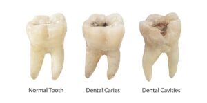
“Dental caries is a microbial disease of the calcified tissues of the teeth characterized by demineralization of the inorganic portion and destruction of the organic substance of the tooth.
History:
Dental caries has its origins rooted deep in history. Even as early as 460 BC to 377 BC, toothache had been described as the worst of tortures. Theories put forward to explain the etiology of dental caries are:
1. Worm theory:
According to an Assyrian legend, tooth ache was caused by worm infestation, which drank the blood of teeth and fed on their roots; as punishment for offences commited against God.
2. Humors theory:
The ancient greeks considered that a person’s physical and mental constitution was determined by four elemental body fluids – blood, phlegm, black bile and yellow bile. All diseases including caries was explained by an imbalance of these humors.
3. Vital theory:
This theory regarded dental caries as originating within the tooth itself, analogous to bone gangrene.
4. Chemical theory:
Parmly proposed that an unidentified ‘chemical agent’ caused caries. He stated that caries began on enamel surface in locations where food putrefied and acquired sufficient dissolving power.
5. Septic theory:
Erdl described filamentous parasites in the surface membrane of teeth/plaque. Ficinus called them ‘denticolae’.
6. Acidogenic theory:
In 1890, W.D. Miller put forth the ‘Acidogenic theory’ or ‘Chemico-Parasitic theory to explain the occurence of tooth decay. According to this theory, dental caries was a result of decalcification of tooth structure due to acids produced by parasites’ or ‘micro organisms’.
7. Proteolysis theory:
In 1944, Gottlieb proposed the “Proteolysis theory. According to this theory, the protein component of tooth structure is destroyed first, prior to loss of tooth minerals.
8. Proteolysis-Chelation theory:
It was Schatz and Martin (1955) who put forth the ‘Proteolysis-Chelation theory’, which was an extension of the proteolysis theory. The products of proteolysis of tooth substance, acquired pellicle and food by bacterial enzymes acts as a chelating agent which remove calcium ions from the tooth.
9. Autoimmune theory:
Jackson and Burch postulated the “Autoimmune theory” of caries development. According to them, the genetic structure of a person was responsible for caries susceptibility or caries immunity. The most widely accepted theory till date continues to be the acidogenic theory put forth by Miller. This can be best understood by the Keyes triad (1960).
The three factors involved in tooth decay are:
1. Tooth(host)
2. Substrate(fermentable carbohydrate)
3. Microorganisms(bacteria)
The Keyes triad was later modified to include
4. Time
5. Saliva
In 1997, Professor Douglas Bratthall devised the Cariogram model to illustrate the caries risk. Caries risk is expressed as ‘percent chance to avoid caries’. A low percentage, for example 5%, indicates a high caries risk. In contrast, 90% chance indicates a very low caries risk.
Components of the cariogram model include :
Chance:
The chance to avoid new cavities in the near future
Diet:
Frequency of eating as well as contents of diet
Bacteria:
Plaque amount as well as types of bacteria
Susceptibility:
Tooth resistance (fluorides) and saliva characteristics
Circumstances:
Past caries experience and systemic diseases and conditions
1. Dietary factors :
Constituents of diet: carbohydrates, proteins, fats, minerals and vitamins.
Carbohydrates :
Type-monosaccharides, disaccharides or polysaccharides
Amount consumed
Form-refined or coarse
Frequency-‘snacking between meals’
Nature-sticky or easily cleared
Biochemical properties-fermentable/non-fermentable.
2. Microorganisms
Acidogenic-S. mutans, Actinomyces
Aciduric-Lactobacilli
Other microorganisms producing IgA1, proteases.
3. Host factors
Morphology of teeth: Deep fissures are prone to food accumulation and development of caries
Intra-oral variation: The caries susceptibility of teeth varies in the following order:
Lower first molar> upper first molars> upper & lower second molars> second bicuspids> upper incisors> cuspids.
Lower incisors and cuspids are least susceptible to caries
*Irregularities of arch forms: Crowding and malaligned teeth favour development of caries
*Saliva-quantity, viscosity, flow-rate, composition, buffering capacity, etc.
4. Genetic factors
5. Immunological factors
6. Other factors
Time
Normally, saliva is supersaturated with calcium and phosphate with respect to tooth mineral which is similar to a carbonated hydroxyapatite.
Whenever carbohydrates are consumed, oral microorganisms rapidly start their fermentation, producing organic acids. These are lactic acid, acetic acid, formic acid etc., with lactic acid being produced in maximum amount. This leads to a fall in pH of the oral fluids. The supersaturation of oral fluids is no longer maintained under such conditions. The organic acids attack the tooth structure, resulting in loss of tooth minerals specifically calcium and phosphate ions, which leach out from hydroxyapatite. This process is known as demineralization .
After a period of 30 minutes, due to salivary buffering by bicarbonate ions, and ammonia production from salivary proteins, there is an increase in the pH of the oral fluids. The acids are neutralized and the conditions now favour precipitation of calcium and phosphate ions onto the tooth structure. This process is known as remineralization and is hastened if fluoride is present even in a small amount in either plaque fluid or saliva.
Natural sugars like lactose and fructose in milk and fruits have as much potential to cause tooth decay as processed sugars. Today, it is thought that starchy foods like ‘potato chips or popcorn’ are worse than foods like sugar or chocolate. This is because these foods being sticky are cleared or washed out from the oral cavity at a slower rate, prolonging their retention time.
The microorganism which is of primary concern in the pathology of dental caries is streptococcus mutans, a subspecies of Mutans streptococci. This microorganism forms insoluble, sticky extracellular polysaccharides: glucans and dextrans. These polysaccharides help in further colonization and increase the contact of the acids with the tooth structure: leading to further demineralization which ultimately leads to cavitation. Thus, dental caries can be viewed as a dynamic process; one of alternating periods of demineralization and remineralization .
The balance between the caries causing and caries protective factors is very delicate. When repeated attacks of demineralization occur there is a net loss of minerals from the tooth surface and caries results. The surface layer of enamel overlying the lesion remains intact and demineralization occurs primarily in the subsurface layers.
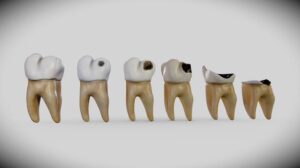
Once caries develops, the process gradually extends deeper, involving enamel and subsequently the pulpo-dentinal organ.
Today, it is also thought that there may be a definite hereditary susceptibility towards dental caries. This may explain why some persons are more prone to decay than other individuals exposed to the same environmental influences. Some investigators have claimed that the caries free and caries active states are two extremes of the spectrum and are determined by a monogenic inheritance pattern, by a single gene. This aspect however has not been proved conclusively. Thus, dental caries, one of the oldest existing and severely crippling diseases known to mankind, awaits research to completely explain its etiology and pathogenesis.
The carious process can be viewed as a complex. multifactorial interaction between the tooth. oral microorganisms and fermentable carbohydrates, with factors like saliva and fluoride playing key roles in this dynamic interplay.
Classification of Dental Caries :
1. Based on location of the lesion :
Pit and fissure caries
Smooth surface caries
Root caries
2. Based on the tissues involved :
Enamel caries
Dentinal caries
Cemental caries
3. Based on the virginity of the lesion :
Primary caries
Secondary caries
4. Based on the progression of the lesion :
Progressive caries Arrested caries
Progressive caries is further classified as:
*Rapidly progressive
-Nursing caries
-Radiation caries
*Slowly progressive
A) Site
Site 1: Includes lesions of pits and fissures on occlusal surfaces of the posterior teeth. These include the buccal grooves of the mandibular molars, palatal grooves of the maxillary molars
Site 2: Includes lesions in the contact areas of posterior and anterior teeth.
Site 3: Includes lesions originating in the gingival third of all teeth.
B) Size
Size 1 (mild): includes lesions which have progressed just beyond remineralization.
Size 2 (moderate): includes larger lesions, with adequate tooth structure to support the restoration.
Size 3 (enlarged): includes lesions in which the tooth structure and the restoration are susceptible to fracture.
Size 4 (severe): includes lesions which have destroyed a major portion of the tooth structure.
Pathogenesis of Dental Caries :
Microorganisms cannot be retained on a clean tooth surface. A biofilm formation is a dynamic process and attachment, growth, removal and reattachment of bacteria is continuous.
Stages in development of dental plaque are :
1. Adsorption of polymers from gingival crevicular fluid.salivary proteins and bacterial cell surfaces to the tooth to form a surface conditioning film, acquired pellicle.
2. Passive transport of microorganisms to the pellicle-coated tooth facilitated by flow of oral secretions.
3. Physiochemical interactions between the microbial cell surface and the pellicle which helps the microorganisms to adhere to the pellicle coated tooth surface.
4. Coaggregation of microorganisms to the already attached cells. This increases the diversity of the microflora mainly late colonizers such as streptococci and actinomyces. 5. Multiplication of attached organisms to produce confluent growth and biofilm formation.
6. Detachment of cells from the biofilm into the saliva. facilitating colonization of fresh sites.
The bacteria produce acids by metabolizing fermentable carbohydrates. causing demineralization. This occurs below the surface zone of enamel causing subsurface demineralization. Clinically, this appears as a white spot lesion because the loss of tooth mineral changes the refractive index.compared with the normal enamel. When sub- surface demineralization progresses to such an extent that the surface layer loses all support, cavitation occurs. During the course of this process, remineralization occurs. Saliva which is supersaturated with calcium and phosphate provides a driving force for remineralization to occur. Partially dissolved crystals act as nucleators for new crystals to grow.
Fluoride hastens this process, leading to formation of fluorapatite which has low solubility and the new mineral is one of low solubility, and is like fluorapatite (fluoridated hydroxyapatite). Fluoride had been postulated to interfere with plaque bacteria in the form of HF i.e. hydrofluoric acid. It was once believed that fluoride available during tooth development altered the morphology of teeth. The molars formed in the presence of fluoride were claimed to have shallower inclines, favorable fissure approximation and more rounded cusps. However, recent evidence suggests that resistance to caries derived through effect on tooth morphology is unsupported and inconclusive.
Progression and Sequelae of Dental Caries :
Once caries is initiated, further progression is inevitable unless controlled by a change in the environment. In enamel. caries starts as a slight demineralization, which leads to loss of translucency. The early enamel lesion consists of four zones with varying levels of demineralization.
1. Transluscent zone
2. Dark zone
3. Body of the lesion
4. Surface zone
As subsurface demineralization progresses towards the dentin, there is further loss of enamel. When the lesion reaches the dentino-enamel junction, the underlying dentine is affected, and there is collapse of the enamel layer leading to obvious cavitation.
Zones of dentin demineralization:
1. Decomposition
2. Bacterial invasion
3. Demineralization
4. Dentinal sclerosis
5. Fatty degeneration
The outer layers of the dentin are heavily infected by bacteria. Both organic matrix and minerals are lost and the dentin is then beyond repair. In the deeper layers, the dentin is affected by plaque acids and demineralisation starts. The number of colony forming units (CFU) of bacteria decreases (about 100 times)in affected dentin. The damage in this layer is reversible if bacterial metabolism can be halted. A barrier of transluscent (well mineralised) dentin may be formed ahead of this layers. Reactionary (secondary) dentin forms to protect the pulp from acid irritation .
If not treated, the lesion progresses towards the pulp. The moment the microorganisms invade the pulp, a carious exposure results. However, bacteria can be preceded by their toxins which can give rise to an inflammatory response in the pulp long before an exposure actually results.
The dental pulp is bound within the confines of rigid dentin. It consists mainly of collagen and absence of collateral circulation. Hence, pressure changes, which occur in the dental pulp(due to inflammation) are so destructive. Chronic pulpal inflammation leads to pulpal degeneration and necrosis. When the necrotic products leave via the apical foramen and reach the periapical tissues. periapical inflammation results, leading to formation of periapical abscess. Subsequent suppuration may ensue and a draining sinus tract may be found intraorally near the gingiva of the affected tooth. This may either resolve or the infection may spread along various tissue spaces.
This generally results in cellulitis . Frequently these changes occur in an insiduous manner and the tooth becomes nonvital without the patient complaining of any symptoms. Cellulitis can lead to potential life threatening situations like cavernous sinus thrombosis, osteomyelitis, Ludwigs angina etc.
Conclusion
It is important to understand the etiological factors related to dental caries in order to prevent the disease before it starts or at least to prevent its progression and complications.