It is the physiologic wearing away of teeth because of tooth to tooth contact, as in mastication. It plays an important physiological role as it helps to maintain an advantageous crown-root ratio and gains intercoronal space of 1 cm, which facilitates third molar eruption.
TYPES
-Physiological attrition-attrition which occurs due to normal aging process, due to mastication.
-Pathological attrition-it occurs due to cartin abnormalities in occlusion, chewing pattern or due to some structural defects in teeth.
ETIOLOGY FACTORS FOR PATHOLOGICAL ATTRITION :-
-Abnormal occlusion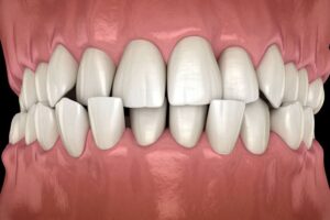
•Development-malocclusion and crowding of teeth, may lead to traumatic contact during chewing, which may lead to more tooth wear.
Acquired- due to extraction of teeth. Extraction causes increased occlusal load on the remaining teeth, as the chewing force for the individual remains constant.
• Abnormal chewing habits-
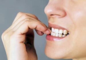 parafunctional chewing habit like bruxism and chronic persistent chewing of coarse and abrasive food or other substances like tobacco.
Occupation-
in certain occupations, workers are exposed to an atmosphere of abrasive dust and can-not avoid getting it into mouth.
•Salivary factor can also play role in attrition.
•Structural defect-in defects like amelogenesis imperfecta and dentinogenesis imperfecta, hardness of enamel and dentin is reduced and such teeth become more prone to attrition.
CLINICAL FEATURES
• Sex-men usually exhibit more severe attrition than women due to greater masticatory force.
• Sites
It may be seen in deciduous as well as permanent dentition.
• It occurs only on occlusal, incisal and proximal surfaces of teeth.
•Severe attrition is seldom seen in primary teeth. as they are not retained for any great period.
• Lingual cusps of maxillary teeth and buccal cusps of lower posterior teeth show most wear.
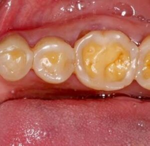
▪︎Appearance-the first clinical manifestation of attrition is the appearance of small polished facet on a cusp tip or ridge and slight flattening of an incisal edge.
Physiological attrition :-
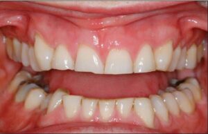
•Physiological attrition begins with wearing of the incisal edge of an incisor, which is followed by the palatal cusp of maxillary molars and buccal cusp of mandibular molars.
•It commences at the time of contact or occlusion.
•Physiological tooth surface loss results in a reduction, in both (vertical) tooth height and (horizontal) tooth width.
Contact points due to slight mobility of teeth in their socket (which is a manifestation of resiliency of periodontal ligament) similar facets occur at contact points.
Color of teeth-when the dentin gets exposed it generally becomes discolored, i.e. brown in color.
•Signs
•There is gradual reduction in cusp height and consequent flattening of occlusal inclined plane.
•There is shortening of the length of dental arch, due to reduction in the mesiodistal diameter of teeth.
•Secondary dentin deposition occurs.
Pathological attrition-
•If pathologically vertical dimension of tooth has reduced then, there is the possibility of com- pensatory growth (dentoalveolar compensation) to some degree.
• Dentoalveolar compensation-if attrition affecting the occlusal surfaces of teeth has occurred, then reduction in occlusal face height (vertical dimension of occlusion) and increase in the freeway space could be anticipated. This may be further complicated by forward posturing of mandible. It is often observed, however, that despite overall tooth surface loss, the freeway space and the resting facial height appear to remain unaltered primarily because of dentoalveolar compensation.
•This is important with respect to patient assessment. If restoration of worn teeth is being planned then the extent of dento-alveolar compensation would appear to determine the dentist’s strategy, defining the need to carry out measures such as crown lengthening, to ensure the same vertical dimension of occlusion and freeway space.
RADIOGRAPHIC FEATURES
•Crown-smooth wearing of incisal and occlusal surfaces of involved teeth is evident by shortened crown image.
•Pulp-sclerosis of pulp chamber and canals is seen due to deposition of secondary dentin which narrows the pulp canals.
• Periodontal ligament-widening of periodontal ligament space and hypercementosis
• Alveolar bone-some loss of alveolar bone.
Differential Diagnosis :-
• Not difficult as it is based on history, location and extent.
Management :-
Treatment of patient depends upon degree of wear relative to the age of patient, etiology, symptoms and patient’s desire.
•Patient should be advised of any possible bruxist habits. The provision of one of three different sorts of splints could be considered. A soft bite guard can help in breaking a bruxist habit or simply will protect the teeth during the bruxist habit. A localized occlusal interference splint is designed to break the bruxist habit and can be wore easily during the day. A stabilization splint reduces bruxism by providing an ideal occlusion: it also enables the clinician to locate and record centric relation. In case of bruxism, use of night guards may be effective in reducing attrition.
•Correction of malocclusion, stoppage of tobacco
chewing habit and restriction of diet to non-coarse food are useful in avoiding attrition.
• Non-carious loss of tooth tissue may require treatment for sensitivity, esthetics, function and space loss in the vertical dimension.