MODIFIED WIDMAN FLAP
In 1965, Morris* revived a technique described early in the twentieth century in the periodontal literature; he called it the “unrepositioned mucoperiosteal flap.” Essentially, the same procedure was presented in 1974 by Ramfjord and Nissle, who called it the “modified Widman flap” . This technique offers the possibility of establishing an intimate postoperative adaptation of healthy collagenous connective tissue to tooth surfaces, and provides access for adequate instrumentation of the root surfaces and immediate closure of the area. The following steps outline the modified Widman flap technique:
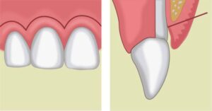
Step 1: The initial incision is an internal bevel incision to the alveolar crest starting 0.5 to 1 mm away from the gingival margin . Scalloping follows the gingival margin. Care should be taken to insert the blade in such a way that the papilla is left with a thickness similar to that of the remaining facial flap. Vertical relaxing incisions are usually not needed.
Step 2: The gingiva is reflected with a periosteal elevator .
Step 3: A crevicular incision is made from the bottom of the pocket to the bone, circumscribing the triangular wedge of tissue containing the pocket lining.
Step 4: After the flap is reflected, a third incision is made in the interdental spaces coronal to the bone with a curette or an interproximal knife, and the gingival
collar is removed .
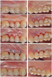
Step 5: Tissue tags and granulation tissue are removed with a curette. The root surfaces are checked, then scaled and planed if needed. Residual periodontal fibers attached to the tooth surface should not be disturbed.
Step 6: Bone architecture is not corrected except if it prevents good tissue adaptation to the necks of the teeth. Every effort is made to adapt the facial and lingual interproximal tissue adjacent to each other in such a way that no interproximal bone remains exposed at the time of suturing. The flaps may be thinned to allow for close adaptation of the gingiva around the entire circumference of the tooth and to each other interproximally.
Step 7: Interrupted direct sutures are placed in each interdental space and covered with tetracycline (Achromycin) ointment and with a periodontal surgical pack.
Ramfjord and Nissle” performed an extensive longitudinal study comparing the Widman procedure, as modified by them, with the curettage technique and the pocket elimination methods that include bone contouring when needed. The patients were assigned randomly to one of the techniques, and results were analyzed yearly up to 7 years after therapy. They reported approximately similar results with the three methods tested. Pocket depth was initially similar for all methods but was maintained at shallower levels with the Widman flap; the attachment level remained higher with the Widman flap.