It is the most common type of pathologic radiolucency encountered in dentistry.
It is a growth of granulation tissue continuous with the periodontal ligament resulting from the death of the pulp and diffusion of bacteria and bacterial toxins from the root canals into the surrounding periradicular tissues through the apical and lateral foramina
Etiopathogenesis :-
It occurs as a response to intense and prolonged irritation from infected root canals producing extension of chronic apical periodontitis beyond the periodontal ligament.
The expanding inflammation and increased vascular pressure result in abscess formation and resorption of the bone in the affected area, which in cause of time is replaced by granulation tissue.
It is the result of a successful attempt by the periapical tissues to neutralize and confine the irritating toxic product that is escaping from the root canal. But continuous discharge into the periapical tissues induce a vascular inflammatory response.
Insult from diseased pulp represents broad spectrum of inflammatory mediations like prostaglandins, kinin and endotoxins.
Elevated level of IgG in pulpoperiapical lesion.
Clinical Features :-
Vitality test-The tooth is nonvital, i.e. it does not respond to thermal and electric pulp test.
Signs-tooth may be darker in color, because of the blood pigments that diffuse into the dentinal tubules.
Symptoms-mild pain can be occasionally experienced while biting or chewing on solid foods.
Sensitivity occurs due to hyperemia, edema and inflammation of the apical periodontal ligament.
There is, seldom, swelling or expansion of the overlying cortical bone. Tooth may feel to be slightly elongated in the socket.
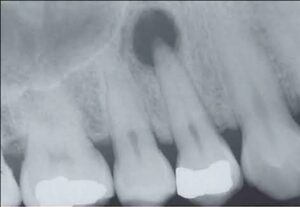
Radiographic Features :-
Lamina dura-periapical area is radiolucent with loss of lamina dura.
• Size-radiolucency is less than 1.5 cm in diameter.
Margins-there may or may not be hyperostotic borders. It may or may not have well defined borders.
Involved tooth may show a deep restoration, extensive caries, fracture or a narrow pulp canal with non-vital pulp.
Periapical rarefying osteitis is the term used to describe the decrease density of loss of lamina dura and periapical bone.
Histopathological Features :-
It consists of proliferating endothelial cells capillaries, young fibroblasts minimum amount of collagen and occasionally, nests of odontogenic epithelium, Rusel’s bodies, foam, cells and cholesterol clefts.
• New capillaries are lined by swollen endothelial cells.
•Types of granuloma according to histopathology
Exudative
Granulomatous
Granulofibrosis
Fibrous
• More inflammation in the center with fibrosis at the periphery.
•Connective tissue is more prominent on the periphery and the bundles of collagen become condensed there, apparently as a result of the slow expansion of the soft tissue mass, resulting in formation of a continuous capsule separating the granulation tissue from the bone.
•Lymphocytes, plasma cells, macrophages, and foreign body giant cells may also be present.
•Occasionally, cholesterol clefts may form the major portion; then it is called as cholesterol granuloma of the jaw.
Differential Diagnosis :-
•Osteolytic stage of cementoma-in dental granuloma tooth is non-vital.
•Surgical defect or periapical scar-tooth shows root canals filling.
Management :-
Extraction of the involved tooth or under certain conditions, root canal therapy, with or without subsequent apicoectomy are the treatment options.