The mucocele is a common lesion of the oral mucosa involving salivary glands and their ducts. They result from traumatic severance of a salivary duct, such as that produced by biting the lips or check, pinching the lip by extraction forceps, and the like, leading to spillage of mucin into the surrounding tissues. Even though such trauma has been implicated in the formation of mucoceles, there is no known history of trauma in many cases. As these cysts lack an epithelial lining, they are not true cysts.
Clinical Features.
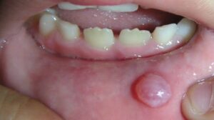
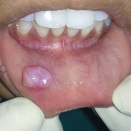
Mucoceles are most commonly found on the lower lip, accounting for over 60 per cent of the all cases and are usually found lateral to the midline. Less common sites include buccal mucosa, anterior ventral tongue (involving the glands of Blandin-Nuhn), and floor of the mouth (ranula).
It is interesting and significant that the mucoceles are restricted almost entirely to the lower lip, seldom found on the upper lip, while accessory salivary gland neoplasms of the lips are almost universally found on the upper lip and only rarely on the lower lip.
Mucoceles are commonly observed in all decades of life, with increased predilection in children and young adults, possibly because of the higher chance of trauma in the latter age group.
Clinically, mucoceles appear as raised, dome- shaped vesicles, ranging in size from 1 or 2 mm to several centimeters. There might be a history of rupture, collapse, and refilling which may be repeated. The mucoceles may lie fairly deep in the tissue or be exceptionally superficial and thus, depending upon the location, will present a variable clinical appearance . Superficial lesions present with a bluish, translucent cast, the blue color imparted by the spilled mucin below the mucosal surface. The deeper lesions are of normal color because of the thickness of the overlying tissue.
Mucoceles often arise within a few days, reach a certain size, and may persist as such for months unless treated. If the contents of the cyst are liberated, they usually are found to consist of a thick, mucinous material. Some lesions regress and enlarge periodically and may disappear after traumatic injury which results in their evacuation. They almost invariably recur, however.
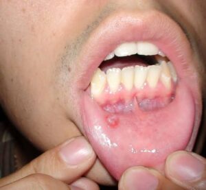
Superficial mucocele, a variant of mucocele, is commonly seen in soft palate, retromolar area, and posterior buccal mucosa, presenting as single or multiple tense vesicles measuring 1-4 mm in diameter. The vesicular appearance is created by the superficial nature of mucin spillage, resulting in a separation of epithelium from the underlying connective tissue. These vesicles often rupture, leaving shallow, painful ulcers that heal within few days. Recurrences are not uncommon in the same location. In some cases, the develop- ment of these lesions may be related to mealtimes. This vesicular appearing mucocele should be differentiated from vesiculobullous lesions, especial- by cicatricial pemphigoid.
Histologic Features.
Mucoceles consist of a circumscribed cavity in the connective tissue and sub- mucosa, producing an obvious elevation of the mucosa with thinning of the epithelium as though it were stretched. The wall of the cavity is made up of a lining of compressed fibrous connective tissue and fibroblasts. Sometimes these cells may be mistaken for flattened epithelial cells. Not uncommonly, the connective tissue wall is essentially granulation tissue, but in any event it usually shows infiltration by abundant numbers of polymorphonuclear leukocytes, lymphocytes, and plasma cells. The lumen of the cyst-like cavity is filled with the spilled mucin containing variable numbers of cells, chiefly leukocytes and foamy histiocytes (macrophages).
Occasional mucoceles demonstrate an intact, flattened epithelial lining. It is probable that this simply represents the portion of the excretory duct bordering the line of severance, if severance is actually the manner in which these lesions develop. The flattened epithelial lining has been referred to as other instances, the epitheliumlined mucocele epithelium of the ‘feeder duct’. In represents a mucous retention cyst.
The salivary gland acini, which lie adjacent to the area of the mucocele and are associated with the involved duct, often show alterations. These may consist of interstitial inflammation or sialadenitis, dilatation of intralobular and interlob- ular ducts with collection of mucus, and break- down of individual acinar mucous cells resulting in the formation of tiny areas of pooled mucus.
Treatment and Prognosis.
Treatment of the mucous retention phenomenon is excision. If the lesion is simply incised, its contents will be evacu- ated, but it will be rapidly filled again as soon as the incision heals. There is occasional recurrence after excision, but this possibility is less likely if the associated salivary gland acini are removed also. The excised tissue should be submitted for microscopic examination to confirm the diagnosis and to rule out the possibility of a salivary gland tumor.