Dental Plaque is an adherent intercellular matrix consisting primarily of proliferating microorganisms, along with a scattering of epithelial cells, leukocytes and macrophages.
Plaque
Plaque can also be defined as the soft deposits that form the biofilm adhering to the tooth surface or other hard surfaces in the oral cavity, including removable and fixed restorations. Dental Plaque is a host-associated biofilm.
Biofilms
Biofilms are defined as “Matrix-enclosed bacterial populations adherent to each other and or/to surface or interfaces (by Costerton). According to the recent data (Widerer and Charaklis 1989) Biofilm is defined as the relatively undefinable microbial community associated with a tooth surface or any other hard, non-shedding material.
DENTAL PLAQUE AS A BIOFILM
Structurally dental plaque is now considered to be a biofilm of complex and dynamic microbial community. It contains areas of high and low bacterial biomass interlaced with aqueous channels of different size, which are the nutrient channels for bacterial colonization. The intercellular matrix forms a hydrated gel in which bacteria can survive and proliferate. Hence, biofilm adheres firmly to the tooth surface and is resistant to mechanical removal, as well as antibiotics.
TYPES OF DENTAL PLAQUE
Based on its relationship to the gingival margin, plaque is differentiated into two categories, supragingival and subgingival plaque
Supragingival plaque is further differentiated into:
Coronal plaque, which is in contact with only the tooth surface, and Marginal plaque, which is associated / with the tooth surface at the gingival margin,
Subgingival plaque can be further differentiated into:
Attached plaque
Unattached subgingival plaque
Attached plaque can be tooth, epithelium and/or connective tissue associated.
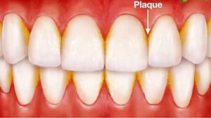
Supragingival Plaque :
It can be detected clinically only after it has reached a certain thickness. Small amounts of plaque can be visualized by using disclosing agents. The color varies from grey to yellowish-grey to yellow. The rate of formation and location of plaque vary among individuals and is influenced by diet, age, salivary factors, oral hygiene, tooth alignment, systemic diseases and host factors.
Subgingival Plaque :
It is usually thin, contained within the gingival sulci or periodontal pocket and thus cannot be detected by direct observation. Its presence can be identified only by running the end of a probe around gingival margin .
Tooth-associated Subgingival Plaque :
The structure is similar to the supragingival plaque. The flora is dominated by Gram-positive cocci, rods, filamentous bacteria and some/few Gram-negative cocci and rods. This flora is associated with calculus formation. root caries and root resorption.
Epithelium-associated Subgingival Plaque :
This type of plaque is loosely adherent because it lacks the interbacterial matrix and is in direct association with the gingival epithelium, extending from the gingival margin to the junctional epithelium. This plaque predominantly contains Gram-negative rods and cocci, as well as a large number of flagellated bacteria and Spirochetes.
Connective Tissue-associated Plaque :
It is usually demonstrated in ANUG and localized juvenile periodontitis patients. The clinical significance is unclear. The unattached plaque can be seen anywhere. Thus, the tooth-associated subgingival plaque is most important in calculus formation, root caries and slowly progressive periodontal destruction, whereas unattached bacterial component is associated with rapid periodontal destruction.
Composition of Dental Plaque:
Bacteria + Intercellular matrix = Dental plaque
Bacteria make up approximately 70 to 80 percent of total material. One mg of dental plaque is estimated to contain 250 million bacteria. Other than bacteria, mycoplasma, fungi, protozoa and viruses may be present. The material among the bacteria in dental plaque is termed as inter microbial/cellular matrix. It contains organic and inorganic portions. The organic matrix is composed of protein-polysaccharide complex produced by micro-organisms.Carbohydrates in the form of levans (fructans) provides mainly energy while glucans (dextran) provide not only energy, but also act as the organic skeleton of plaque. Other carbohydrates are galactose and rhamnose. Glycoproteins/provide the protein component and small amounts of lipids are also present. Inorganic components include, primarily calcium, phosphorus with small amounts of magnesium, potassium and sodium.
FORMATION/DEVELOPMENT OF DENTAL PLAQUE
Pellicle is the initial organic structure that forms on the surfaces of the teeth and artificial prosthesis. The first stage in pellicle formation involves adsorption of salivary proteins to apatite surfaces. This results from the electrostatic ionic interaction between hydroxyapatite surface which has negatively-charged phosphate groups that interacts with opposite charged groups in the salivary macromolecules. The mean pellicle thickness varies from 100 nm at 2 hours to 500 to 1,000 nm.
The transition from pellicle to dental plaque is extremely rapid. The first components include mainly cocci with small number of epithelial cells and PMNL’s they form a monolayer within a few hours, and the attached bacteria proliferate and form small colonies of cocci. With time other types of microorganisms proliferate and form different microcolonies.
Hence, in dental plaque development, two adhesion processes are required. First, bacteria must adhere to the pellicle surface and become sufficiently attached to withstand oral cleansing forces. Second, they must grow and adhere to each other to allow plaque accumulation.
Bacterial Adherence :
During initial adherence, interactions occur mainly between specific bacteria and the pellicle. They are:
Bacterial Attachment via Electrostatic Interactions :
Oral bacteria bear an overall net negative charge, negatively-charged components of the bacterial surface and negatively-charged components of pellicle become linked by cations such as calcium
Bacterial Attachment via Hydrophobic Interactions :
These interactions are based on the close structural fit between molecules on the pellicle and bacterial surfaces. The nature of the hydrophobicity of the cell is not clearly known. The contributing factor might be (LTA) lipoteichoic acid, which may provide a long hydrophobic area
Bacterial Attachment via Specific Lectin-like Substances :
Lectins in the bacterial surfaces recognize specific carbohydrate structure in the pellicle and become linked Adhesion and attachment occurs between:
1. Bacteria and clean tooth surface 2.Bacteria and pellicle
3.Bacteria and same species
4.Bacteria and different specie
5.Bacteria and matrix
Next step in plaque formation is:
Growth and Accumulation of Bacteria
Once the bacteria is adhered to the pellicle, subsequent growth leads to bacterial accumulation and increased plaque mass. Dental plaque growth depends on:
a. Growth via adhesion of new bacteria b. Growth via multiplication of attached bacteria
The initial bacteria that colonize the pellicle surface are mostly Gram-positive facultative microorganisms such as Actinomyces viscosus and Streptococcus sanguis, as the plaque matures, secondary colonization of Prevotella intermedia, Capnocytophaga, Porphyromonas gingivalis takes place. This ability of bacteria to adhere to different species and genera of micro-organisms is known as co- aggregation.