- Dentinal Sclerosis
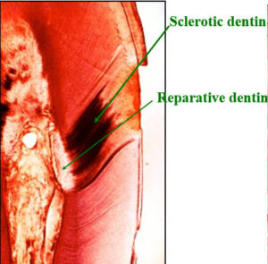
Sclerosis of primary teeth dentin is a regressive alteration in tooth substance that is characterized by calcification of the tooth dentinal tubules. It occurs not only as a result of injury to the teeth dentin by dental caries or abrasion but also as a manifestation of the normal aging process.
For many years it has been known that if a ground section of a tooth with a very shallow dental carious lesion of the tooth dentin is examined by transmitted light, a translucent zone can be seen in the dentine underlying the cavity or dental caries. This was readily recognized as being due to a difference between the refractive indices of the sclerotic or calcified dentinal tubules and the adjacent normal tubules. Both Beust and Fish showed that dyes do not penetrate those dentinal tubules that are sclerotic as a result either of age or of a slowly progressive type of dental caries.
The exact mechanism of dentinal sclerosis or the deposition of calcium salts in the tubules is not understood, although the most likely source of the calcium salts is the fluid or ‘dental lymph’ within the tubules. The increased mineralization of the tooth decreases the conductivity of the odontoblastic processes. In addition, the sclerosis slows an advancing carious process.
Sclerotic dentin under a carious lesion was shown by Hodge and McKay to be actually harder than adjacent normal teeth dentin. Subsequently. Van Huysen, Hodge, and Warren confirmed the fact that sclerotic dentin is more highly calcified than normal dentin by employing a unique adaptation of the dental x-ray absorption technique in which ground slabs of teeth were photographed by x-rays and the degree of radiopacity in areas of normal and sclerotic dentin was measured.
Development of Dentinal Sclerosis
Visible sclerosis of small isolated areas of the dentin commonly be comes evident after the period of adolescence.
Indication of beginning changes may rarely be seen as early as the time of tooth eruption. The exact nature of the modification occurring in the dentin has not yet been accurately determined.
A hypercalcification, possibly combined with a keratinization, appears to occur. The result of the process is a sclerosis of the dentin matrix sub stance, followed by a similar modification of the organic content of the tubules. Sclerosis ultimately results in obliter ation of the tubules; which hinders the passage of metabolic fluids. Observations indicate that it also precludes the en trance of the agents that cause caries.
Sclerosis has, therefore, a marked in fluence on the behavior of the tooth in respect to its environment. Experiments have indicated that resistance of the tooth to caries and trauma increases in propor tion to the amount of sclerosis.
Physiologically, two types of sclerosis may be conveniently differentiated. An irritation sclerosis is a reaction to such trauma as abrasion, erosion or the presence of congenital or traumatic enamel Work supported in part by a grant from the Physiology, Pathology, Bacteriology and Chemistry
A spontaneous sclerosis occurs without visible cause in teeth that are perfect in form and function; or from age changes, such as the maturation process. This may lead to complete hy percalcification of the tubules of the crown and root and finally to calcifica tion of the pulp.
Anatomically, two forms of sclerosis may be distinguished. An opaque form, marked by a predominance of opaque tubules, is the most common. A trans parent form, marked by a relative free dom of opaque tubules, may be an advanced stage of the former. Specimens partaking of the characteristics of both may be conveniently described as mixed. These forms have been considered in recent publications.
As the tubules and matrix substance in sclerosed areas are impervious to stains, the condition can conveniently be demon strated by subjecting whole or halved teeth or ground sections to an intensive staining process. Dentin with normal open tubules is permeable. Such dentin is chromophilic. Sclerosed dentin, on the other hand, is not colorable by ordinary means and is therefore designated as chromophobic. To demonstrate the various degrees of sclerosis in a stained ground section to an audience would require three photographs, one of which must be made by transmitted and two by reflected light. For practical purposes, the major amount of sclerosed tissue found in a section or halved tooth .