About Dr. Gourav Sharma:
![]()
Senior Painless Dental Surgeon in Para Military Hospital
Para Military ITBP Hospital, Indo-Tibetan Border Police Force, Government of India, Shivpuri, Madhya Pradesh
2013 to 2017 Shivpuri, Madhya Pradesh
Modern Painless techniques and Sophisticated medicines in Para Military Hospital.

Apollo Hospital Senior Associate Dental Surgeon
Physical Treatment & Online Dental Consultation to the Patients.
 Lecturer & Dental Surgeon Mansarovar Hospital & Research Center
Lecturer & Dental Surgeon in Mansarovar Hospital and Research Center Bhopal, Madhya Pradesh
- 2011 to 2012
 Senior Painless Dental Surgeon.
2011 to current in own Parthsarathy Dental Clinic Shivpuri, Madhya Pradesh
 Senior Healthcare Consultant at Practo
2021 to current with own clinic online and Physical support to the Patients
- Provide online support to the patients, when the problem does not report as complete or require Dentist assistance.
Developmental Disturbances of Teeth
SIZE OF TEETH
Microdontia
Microdontia refers to teeth that are smaller than normal. There are three types of microdontia.
True generalized-all the teeth are smaller than normal. It occurs in pituitary dwarfism, Down syndrome and congenital heart disease.
Relative generalized-normal or slightly smaller than normal teeth; are present in jaws that are somewhat larger than normal. It is hereditary. It often exhibits spacing between the teeth..
Localized-it involves only single tooth. It occurs with congenital heart diseases, Down syndrome and progeria.
Clinical features
Localized microdontia
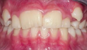
Most commonly affected teeth are maxillary lateral incisors and 3rd molars.
Supernumerary teeth are frequently smaller than normal.
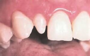
Peg shaped lateral-it is one of the common form of localized microdontia is peg shaped laterals in which the mesial and distal sides converges or taper incisally, forming peg shaped or cone shaped crown.
. Molars when molars are involved, they may also undergo a change in shape from five to four cusps in case of mandibular molar and from four to three cusps in upper molars.
Radiographic features
Radiograph will permit evaluation of size of both, erupted and non erupted teeth.
Management
Crown and bridge work is required for esthetich rehabilitation of teeth.
Macrodontia
It is also called as ‘megadontia’. These are the teetha which are larger than normal. It is of three types:
True generalized-all the teeth are larger than normal. It is commonly associated with pituitary gigantism.
Relative generalized-teeth are normal or slightly larger than normal, but present in a smaller jaw.
Localized-one or more large teeth exist in relation to an otherwise normal dentition and body size.
Causes
It is occasionally seen in facial hemi-hypertrophy, in which half of the teeth in unilateral distribution are affected.
Angioma of face, pituitary gigantism and genetich component.
Clinical features
Teeth size-teeth are larger than normal.
Malocclusion-there is crowding, which may result in malocclusion.
Impaction-as space is less, there is impaction of teeth
It should not be confused with fusion.
Radiographic features
The increased size will be demonstrated on the radiograph.
Management
If necessary orthodontic treatment is done.
If impacted, extraction is indicated.
SHAPE OF TEETH
Gemination
Gemination refers to the process whereby, in a single tooth germ invaginates resulting in incomplete formation of two teeth that may appear as bifid crown on single root. It occurs during the proliferation stage of the growth cycle of tooth.
Etiology
Hereditary and familial tendency.
It results from the splitting of a tooth germ during development or from the fusion of a normal tooth bud with a developing supernumerary tooth.
Clinical features
Sex-males and females are equally affected.
Sites-commonly affected teeth are deciduous mandibular incisors and permanent maxillary incisors.
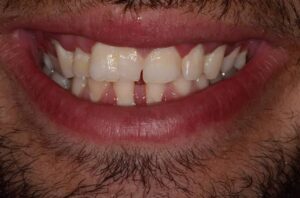
Appearance-it appears clinically as bifid crown with single root.
It does not increase or decrease the number of teeth present.
Crown features
There are common pulp canals and either single or partially divided pulp chambers.
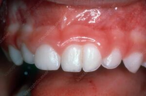
Crown is wider than normal with shallow groove extending from incisal edge to cervical region.
Enamel or dentin of crown of geminated teeth may be hypoplastic or hypocalcified.
Invagination of crown occurs with complete and incomplete division.
Radiographic features
• Altered morphology of geminated tooth’s hard tissue and pulp chamber.
Cleft in the crown and invagination are usually outlined by the radiopaque enamel which accentuates them.
Pulp chamber is single, enlarged and may be partially divided.
Differential diagnosis
Fusion-tooth structures with two separate root canals and with either one root or two roots is result of fusion, in contrast to the enlarged and possibly partially divided pulp chamber in an enlarged tooth with bifid crown which is a result of gemination. In gemination the full complement of tooth is present while in the case of fusion, tooth is missing.
Management
Affected tooth structure should be removed and crown may be restored and reshaped.
Reduction of mesiodistal width with periodic disking.
Jacket crown preparation.
Complication
Pulpal infection-areas of hypoplasia anda invagination lines or areas of coronal separation represent caries susceptible area, which may lead to pulpal infection.
Others-it may also cause malocclusion anda periodontal pathosis.
Twining
Twining indicates cleavage of tooth germ which results in formation of supernumerary teeth that is mirror image or near image of tooth from which it has developed.
Fusion
Fusion is also called as ‘synodontia’. It represents the embryonic union of normally separated tooth germs. It represents junction at the level of dentin between juxtaposed normal tooth germs.
Etiology
Genetic-it is transmitted as autosomal dominant trait with reduced penetration.
Physical-physical force or pressure generated during development causes contact of tooth germs.
Two separate developing tooth germs being initially close together; as they grow and expand; they contact with each other and the germs fuse to varying degrees.
Classification
Complete-if fusion takes place before calcification begins, the two teeth may be completely united to form a single large tooth.
Incomplete-if contact of teeth occurs later, i.e. when the portion of crown has completed its formation; then there is union of root only.
Clinical features
Sex-male to female ratio is 1:1.
Site and area-it is seen more commonly in anterior teeth. It is more common in deciduous dentition than in permanent dentition.
It may occur between a normal tooth and a supernumerary tooth such as ‘mesiodens’ or ‘distomolar’.
Tooth is almost twice in size than normal, with or without bifid crown.
Tooth may have separate or fused root canals.
Clinical problems-it may be related to appearance,
spacing and periodontal conditions.
Significance
Dental caries is common in fused teeth.
It may result in reduced number of teeth in the jaws.
When deciduous teeth fuse, the corresponding permanent teeth may be absent.
Radiographic features
True nature and extent of the union will be more evident on radiograph.
Differential diagnosis
Gemination-it is discussed in differential diagnosis of gemination.
Management
Morphology of teeth should be determined radiographically for endodontic treatment.
After endodontic treatment, tooth may be reshaped with a restoration that will mimic independent crown.
Concrescence
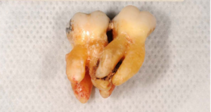
Concrescence is a form of fusion that occurs after the root and other major parts involved in teeth are formed or when the roots of two or more teeth are united by cementum, below the cementoenamel junction. It is also called as ‘false gemination’.
Etiology
Traumatic injury.
Overcrowding of the teeth with resorption and interdental bone loss.
Distal inclination of crown of molar,
Space restriction during development.
Excessive occlusal trauma.
Local infection after development.
Types
True concrescence-if roots are bound during development.
Acquired concrescence-if the condition occurs after development.
Clinical features
Sex-male to female ratio is 1:1.
Site and area-it is common in maxillary 2nd and 3rd molar area. Either primary or secondary teeth are affected.
Usually involved are only two teeth, roots are fused by cementum.
Significance-teeth may fail to erupt or incompletely erupt.
Malocclusion and impaction-there may be malocclusion or the teeth may be impacted.
Radiographic features
Diagnosis is made by radiographs.
It is not always possible to distinguish between concrescence, teeth in close contact and superimposed teeth.
Management
Dentist must be careful while doing extraction.
Talon’s Cusp
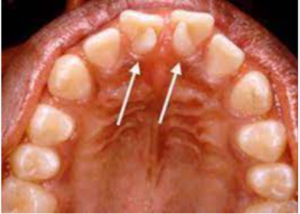
Talon’s cusp projects lingually from cingulum area of maxillary and mandibular teeth or it is an anomalous hyperplasia of cingulum on the lingual of maxillary and mandibular incisors, resulting in the formation of supernumerary cusp
Pathogenesis
A focal proliferation of tissue during development.
Exuberant development of the fourth lobe (cingulum).
Clinical features
Sex and site-it may be found in both sexes anda common in both dentitions.
Appearance-it resembles like an ‘Eagle’s talon’.
Signs-it blends smoothly with the erupted tooth, except that there is deep a developmental groove where the cusp blends with sloping lingual tooth surface.
Composition-it is composed of normal enamel. dentin and contains a form of pulp tissue.
Shape-cusp may or may not contain pulp horn and is usually ‘T shaped.
Significance
Patients face problems with esthetic.
There is also high incidence of caries.
Occlusal interference may be there.
• Syndrome-it is associated with Rubinstein-Taybi syndrome.
Radiographic features
Superimposed with incisors, on which it occurs
Outline is smooth and a layer of normal appearing enamel is distinguishable.
Differential diagnosis
Supernumerary teeth-close association can be determined by tube shift technique.
Management
Prophylactic restoration of the groove.
Removal of cusp followed by endodontic therapy.