Recording the Orientation Jaw Relation using A Kinematic Face-bow in a Dentulous Patient :
The mechanism of operation of the apparatus is similar to arbitrary face-bow. Of course there are a few differences, which are as follows:
• An accurate impression of the mandible and maxilla should be made.
• Stone casts are fabricated using the impres sions.
Two-piece custom-built metal clutches are fabricated .
• A stem is attached to the centre of the labial surface of the clutch. The stem should be parallel to the sagittal plane .
• The cast aluminium clutches (maxillary and mandibular) are attached to the teeth using Zinc oxide eugenol impression paste.
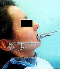
• The face-bow is positioned on the face such that:
The mandibular clutch is attached to the U-frame of the face-bow. The stem is passed through a provision in the U-frame .
The maxillary clutch is attached to the horizontal bar of the face-bow .
• The horizontal arm is fixed such that the grid is placed over the area anterior to the external acoustic meatus
. The metal styli (rods) at the distal end of the face-bow should be adjusted/positioned over the condyle (13 mm in front of the tragus on the cantho-tragal line). The patient should be in a semi-supine position while positioning the styli. The styli should contact the grid of the horizontal arm.
• The patient is asked to slowly open his mouth to a maximum of 20 mm inter-incisal distance. This is very important because only with this range of jaw separation, the mandibular condyles show pure rotation. Beyond this jaw separation, the mandible begins to translate. The mandible shows pure rotation along the hinge axis. The patient should be trained to open and close his mouth at the hinge axis.
• As the patient moves his mandible, the styli will first form an arc on the grid. But once the styli reach the hinge axis, they stop and begin to rotate at that particular point . The grid is gently removed and the styli are made to contact the skin. The point of contact is tatooed on the skin for further reference. This completes the face-bow recordm i.e. the true hinge axis has been located.
Pantographic Tracing :-
• After completing the face-bow record, jaw writing is recorded in three planes. A panto- graph is required to record the jaw writings (mandibular movements recorded on flags using styli) in three planes . Panto- graphs are used to record the mandibular movement and also to develop satisfactory occlusion on an articulator. They record the mandibular movement in relation to an estab- lished plane in the face (a plane formed by the hinge axis along with the anterior reference points).
• A pantograph has two horizontal bars attached to metallic clutches like a kinematic face-bow assembly. But here the distal end of the upper bar has two grids (instead of one) placed perpendicular to one another. Two anterior grids are placed on the lower bar. Each grid has a separate stylus to draw the mandi- bular paths. The metal styli should be oriented to the tatooed hinge axis.
• The patient is made to rehearse hinge move- ment and translatory movement like right lateral and left lateral. A Cohen or Hitchkok trainer can be used to rehearse the mandibular movements.
After training the patient, the grids are coated with a pressure sensitive material (pumice- ether mix). The stylus of each grid is made to contact the grid and the patient is asked to perform the trained movements. The styli will draw tracings on their respective grids when the patient moves his mandible. These tracings are thin and delicate line and are known as pantograms .
• The tracings on the condylar grids will be slightly different.
• These tracings can be used to program a fully adjustable articulator, which is useful to develop the ideal occlusion for a complicated restorative case.
• Occlusion developed by such method will exhibit no posterior tooth contact when the mandible is in any eccentric position. Contact is present only between the cusp tip and the ridges around the central fossa.
Treatment Procedure :-
We know that full mouth rehabilitation is a combination of multiple procedures done to restore the oral cavity to its original condition. The basic steps to be followed while carrying out a complete oral rehabilitation procedure have been enumerated.
Diagnosis and treatment planning.
Diagnostic casts should be accurately moun- ted on an adjustable articulator to study the mandibular movements and premature contacts that occur during eccentric movements.
• Three sets of impressions are necessary
One, for diagnosis and permanent record.
Second, for the construction of clutches.
Third, for mock preparation. This includes preoperative estimation of tooth reduction and development of articulation (dynamic occlusion) in wax.
After face-bow transfer using an arbitrary facebow, the first set of casts (study casts) are mounted using the centric relation record using split-cast technique (casts are indexed with grooves so that they can be easily remounted).
A pair of clutches are fabricated using the second set of casts to record the true hinge axis (THA) of the patient using a kinematic facebow.
The articulators (semi and fully adjustable) are reprogrammed according to the THA.
• The study casts are remounted according to the programming done on the articulator.
• After locating the hinge axis, the plane of orientation of the maxillary ridge (orientation jaw relation) is transferred to the articulator using a conventional face-bow. The THA on either side form the first two reference points. The anterior reference point on the face is used as the third point of reference to orient the plane of the maxilla .
The face-bow record is transferred to the programmed articulator and the maxillary cast is mounted.
• Next, the centric relation record is made and used to mount the mandibular cast against the maxillary cast.
Now the casts are in opening and closing relation to the terminal hinge position. But this is not sufficient to reproduce all the mandibular movements. To reproduce other movements of the mandible in the articulator, special instruments can be used. (Note: The casts should be accurately mounted in relation to the opening axis of the articulator. The relationship of the casts to the opening axis should be a duplicate of the relationship between the temporomandibular joint and the arches in the patient. This is crucial to develop harmonious occlusion during oral rehabilitation).
• The centres of lateral movement should be located using a Twin Gothic Arch Tracing (needle point). After locating THA and the centre of lateral motion, the paths of these centres are recorded using a pantograph. The articulator is further programmed according to the pantographic tracings (pantograms). Method of recording a pantographic tracing has been described alongwith kinematic facebow.
Finally, the casts are studied and an appropriate treatment plan is drawn up depending on the requirement of the patient.