LIPOMA
It seldom occurs in oral cavity. It is a benign, slow growing, tumor composed of mature fat cells.
Types
Encapsulated lipoma-it is the commonest tumor.
Diffuse lipoma it does not posses typical features of lipoma. It is also called as ‘pseudolipoma’.
Lipomatosis it has multiple lipomas. It refers to the symmetrical masses of fat, which sometimes occur around the neck in middle age man and as painful deposits of fat in women in Dercum’s disease.
Clinical Features
Age and sex distribution-occurs after 40 years with peak at 50 years; male to female ratio is 1:1.
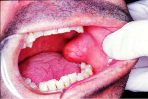
Common sites-it usually occurs in upper parts of the trunk, neck and arms. In oral cavity, it occurs on buccal mucosa and mucobuccal fold followed by tongue, floor of mouth and lip.
Appearance-it appears as a solitary lesion with sessile, pedunculated or submerged base.
Size-size of lesion is approximately 1 cm in diameter.
Margins- well contoured, well defined, round to large ill defined lobulated mass.
Shape-it grows as round or ovoid mass in oral cavity.
Base-it may be lobulated or may be broadly based or have narrow pedicle.
Color-due to thinness of the overlying epithelium, yellow coloration of the fat can be seen.
Signs
Most of the lesions are fluctuant and are not freely movable.
Surface is smooth, non tender, soft and cheesy in consistency.
The epithelium is usually thin and the superficial blood vessels are readily visible over the surface.
Some lesions of lipoma are deep and feel fluid on palpation; may be mistaken as cysts.
Slip sign the edge of lipoma is soft, compressi- ble and often slips away from the examining fingers.
Transillumination test-it may be positive
Lipoblastomatosis-it is a variant of lipoma, but is not a true neoplasm. It is the continuation of the normal process of fetal fat development carried into the postnatal life. It is characterized clinically by the occurrence in infants of solitary or multiple soft tissue masses, developing at various sites such as the buttocks, chest, axilla or neck.
Hibernoma-it develop as multi-vacuolated fat that is analogous to the brown fat of hibernating ani- mals; however lesions in oral cavity are not reported.
Histopathological Features
It is made of circumscribed mass of mature fat cells, which may exhibit varying number of collagen strands coursing through the lesion and supporting occasional small blood vessels.
Nucleus is flattened against the cell wall, as it is filled with fat.
The tumor cells are arranged in lobules with an intervening connective tissue stroma.
Management
Surgical excision and recurrence is uncommon.