It is a condition which was first described by Ludwig in 1936. The word angina means sensation of choking or suffocation. It is the most commonly encountered neck space infection.
This condition may be defined an overwhelming, rapidly spreading, septic cellulitis involving submandibular, submental and sublingual spaces bilaterally.
Etiology :-
Odontogenic infection-it is usually an extension of odontogenic infection from mandibular molar teeth into the floor of mouth. The 2nd and 3rd molars are the teeth most commonly involved.
Trauma-it may also be caused by oral soft tissue laceration and punctured wounds of the oral floor.
Sialadenitis-submandibular gland sialadenitis and infected malignancy may be sometimes contributory to Ludwig’s angina.
Calculi-salivary calculi or from intravenous injection of the internal jugular vein, especially in drug abusers.
Osteomyelitis-osteomyelitis mandibular fracture.
Bacteriology :-
Streptococci are the most commonly reported organism from the culture. .
Other microorganisms are a-hemolytic streptococci, Bacteroides, Klebsiella, Fusiform bacilli oand E. coli.
Clinical Features :-
There are three typical appearances of Ludwig’s angina
First
It is characterized by brawny indurations. Tissues are board like and do not pit on pressure. No fluctuance is present.
The tissues may become gangrenous and when cut, they have a peculiar lifeless appearance.
A sharp limitation is present between the infected tissues and surrounding normal tissues.
Second
Three facial spaces are involved bilaterally, i.e. submandibular, submental and sublingual. If the involvement is not bilateral, the infection is not considered a typical Ludwig’s angina .
Third
• The mouth is open and the tongue is lifted upwards and backwards, so that it is pushed against the roof of the mouth and the posterior pharyngeal wall; when this occurs, acute respiratory obstruction is likely to occur.
• Two large potential spaces at the base of the tongue are involved, i.e. submental and sublingual.
Other features :-
Signs
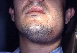
Swelling is firm, painful and diffused, with no sign of localization.
Floor of mouth appears erythematous and edematous. Stiffness in tongue movement generally develops.
The patient develops a toxic condition and speech becomes impaired.
Larynx and glottis become edematous.
• As the disease continues the swelling starts involving the neck.
Symptoms :-
The patient always has fever and there is
considerable salivation, as the patient is unable to swallow. There are chills accompanied with fever.
There is an inability to swallow and to eat. Respiratory distress is common..
There is also neck pain, redness of neck, fever, weakness, fatigue, excessive tiredness, confusion or other mental changes, difficult breathing and earache.
There is an intense pain on tongue movement and the patient may be severely dehydrated, owing to inability to take anything by mouth.
•If the swelling has spread into the pterygoid region, then there is difficulty in opening the mouth.
Fatal Complications :-
• Respiratory obstruction-the infection of Ludwig’s angina tends to spread through the connective tissues which cover the small muscles of the larynx and between the muscles of the floor of mouth. The epiglottis and larynx become edematous along with the posterior aspect of the tongue. The tongue gets elevated and gets pressed upward and backward, causing pressure on the larynx. Therefore, dyspnoea occurs in paroxysm. Ultimately, death occurs due to respiratory embarrassment.
Generalized septicemia.
Erosion of the carotid artery-late spread of infection to lateral pharyngeal space can also cause erosion of the carotid artery.
Cavernous sinus thrombosis with subsequent meningitis may be sequelae of it..
• Others mediastinum extension, pharyngomaxillary space extension, osteomyelitis and airway obstruction.
Prevention :-
Regular visits to the dentist and prompt treatment of oral/dental infections can prevent the conditions that increase the risk of developing Ludwig’s angina.
Laboratory Findings :-
A moderate leukocytosis is found.
Fusiform bacilli and spiral forms, various staphylo- cocci, diphtheria and may other microorganisms have been cultured on different occasions.
Management :-
Intense and prolonged antibiotic therapy penicillin is to be administered IM or IV, in high doses, because it is the empirical antibiotic of choice and the oral flora, including most of anaerobes, are sensitive to it. Recently, combination of gentamicin and cloxacillin has been proved successful.
• Establishment and maintenance of an adequate airway are the essentials of therapy-tracheostomy is a routine procedure, but it is often difficult to perform in the late stages. Attempt at blind intubations is often time consuming. Recently, successful intubations with fibreoptic laryngoscope have been introduced as a worthwhile technique in the therapy of Ludwig’s angina. In the late stage, cricothyroidotomy may be performed as an emergency procedure.
Incision and drainage-it is done for the release of tissue tension. A horizontal incision, midway between the chin and hyoid bone, is a classic approach to surgical drainage of Ludwig’s angina. It may be carried out under local anesthesia.
• Supportive therapy-parenteral hydration, high protein diet and vitamin supplements.
Extraction of offending tooth.