PERIAPICAL ABSCESS :
(Dento-alveolar abscess, Alveolar abscess)
The periapical abscess in an acute or chronic suppurative process of the dental periapical region. It may develop either from acute periapical periodontitis or more commonly from a periapical granuloma. Acute exacerbation of a chronic peri- apical lesion is also called a Phoenix abscess.
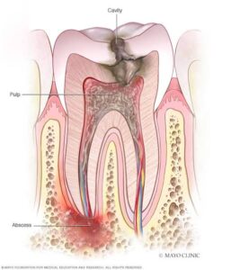
It usually arises as a result of infection following car- ious involvement of the tooth and pulp infection, but it also does occur after traumatic injury to the teeth, resulting in necrosis of the pulp, and in cases of irritation of the periapical tissues either by mechanical manipulation or by the application of chemicals in endodontic procedures. It is a mixed infection with the culture of pus yielding to a wide range of different bacterial species.
Clinical Features :
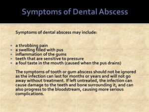
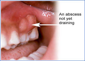
The acute periapical abscess presents the features of an acute inflammation of the apical periodontium. The initial stages produce tenderness of the tooth, which is often relieved by application of pressure. In time, the tooth is extremely painful and is slightly extruded from its socket. As long as this abscess is confined to the immediate periapical region, there are seldom severe systemic manifestations, although regional lymphadenitis and fever may be present. However, rapid extension to adjacent bone marrow spaces frequently occurs, producing an actual osteo- myelitis, but this is sometimes still considered clinically to be a dentoalveolar abscess. In such cases the clinical features may be severe and seri- ous with swelling of the tissues.
The chronic periapical abscess generally presents no clinical features, since it is essentially a mild, well-circumscribed area of suppuration that shows little tendency to spread from the local area .
Roentgenographic Features :
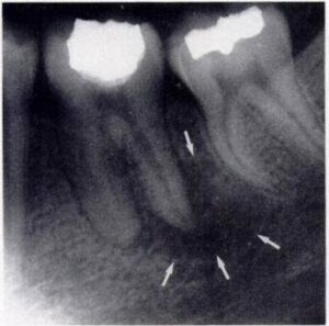
The acute periapi cal abscess is such a fapidly progressive lesion that, except for slight thickening of the periodontal lig ament space, there is usually no roentgenographic evidence of its presence. The chronic abscess, developing in a periapical granuloma, presents the radiolucent area at the apex of the tooth described previously or the radiolucency may be ill-defined
Histologic Features :
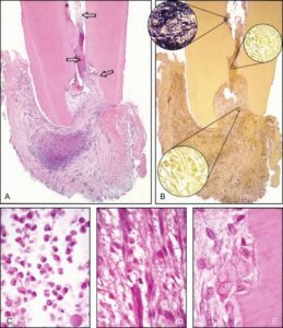
The area of suppuration is composed chiefly of a central area of disintegrating polymorphonuclear leukocytes surrounded by viable leukocytes, occasional lymphocytes, cellular debris, necrotic materials and bacterial colonies. There is dilatation of the blood vessels in the peri- odontal ligament and adjacent marrow spaces of the bone. These marrow spaces also show an inflammatory cell infiltrate The tissue around the area of suppuration contains a serous exudate.
Treatment and Prognosis :
The principle of treatment of the periapical abscess is the same as for any abscess: drainage must be established. This can be accomplished by either opening the pulp cham- ber or extracting the tooth. Under some circum- stances, the tooth may be retained and root canal. therapy carried out if the lesion can be sterilized.
If the periapical abscess is not treated, it may lead to serious complications through the spread of the infection. These include osteomyelitis, cellulitis, and bacteremia and the ultimate formation of the fistulous tract opening on the skin or oral mucosa. Cavernous sinus thrombosis has also been reported.