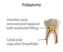
Pulpotomy:
It is defined as the removal of coronal portion of infected vital pulp.
Objective :
To maintain vitality and function of remaining non- infected healthy pulp tissue in root canal.
Amputed surface of pulp again becomes covered with odontoblast which forms a layer reparative dentin protecting the pulp in root canal.
Procedure :
▪︎Obtain profound anaesthesia.
▪︎Isolate the tooth by rubber dam.
▪︎Standard cavity preparation is done by following all steps of cavity preparation.
▪︎Remove roof of pulp chamber, this examination of odontoblastic membrane.
▪︎Amputation of pulp is done.
▪︎Arrest the haemorrhage by inserting a wet sterile pellet into pulp chamber with slight pressure.
▪︎Place the dressing of Ca(OH), suspended in methyl cellulose.
▪︎Protection of dressing is done by apply of ZOE cement base.
▪︎Take radiograph before discharging the patient.,check the occlusion and relieve any high point.
▪︎Call the patient after one week to observe any unlike reaction.
▪︎Final restoration is done at 6th week for posterior silver amalgam and for anterior composite is used.
▪︎Periodic recall visits are set up at every 3 months and vitality is checked and radiograph is taken and is compared with previous one for formation of calcified bridge.
It is done till 3½ years.
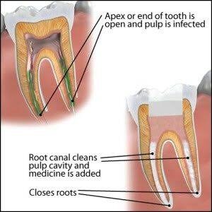
Apexification :
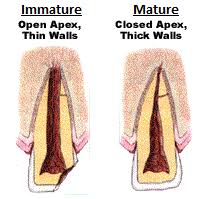
Apexification is a method to induce the development of root apex of an immature pulpless tooth by formation of osteocementum or other bone like tissue.
Objective :
The aim of apexification is to close the open apical third of the root canal or the formation of an apical calcific barrier against which obturation can be achieved.
Technique :
-Obtain anaesthesia.
-Apply rubber dam and isolate.
-Prepare access cavity for biomechanical preparation. Establish the accurate length of root canal with radiograph, the working length should be atleast 2 mm short of root length to prevent injury to periapical tissue and at cervical one third of root.
-Now remove the content of pulp cavity with the help of broaches, flush the root canal with 5.2% sodium hypochloride and H₂O..
-Biomechanical technique enlarge the root canal upto radiographic apex.
-Circumferential filling is done.
-Use large files no. 60-120 are required, try to remove as much necrotic tissue as possible.
-Dry the root canal with paper points.
-Prepare thick paste of Ca (OH), in water or local anaesthetic solution and pack into root canal with plugger.
-Avoid foreign substance beyond open apex, now the opening is sealed with double seal (gutta-percha and ZOE cement outside).
-Check high point and take radiograph.
-Recall after 2-3 days and check for any inconvenient reaction, if no complaint fill the access cavity with silver amalgam in posterior and composite in anterior.
-Recall after 3 month interval, normal time require for apexification is 6 month to 2 years.
-Closure verified on radiograph and clinically by opening root canal and testing with blunt instrument meeting a definitive stop.
-The RCT is done with permanent root canal filling of gutta-percha, for anterior apply porcelain crown. For posterior apply full cast crown.