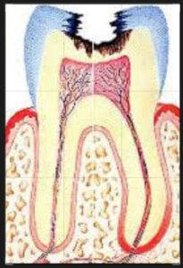
It is a mild to moderate inflammatory condition of the dental pulp of teeth caused by noxious stimuli in which the dental pulp is capable of returning to uninflamed state following removal of the stimuli.
Causes :-
It may be caused by any agent that is capable of injuring the tooth pulp like trauma, disturbed occlusal relationship, and thermal shock.
Pathogenesis :-
• Some stimuli of short duration, such as cutting dentin of teeth may cause short-term vasodilation and a reversible increase in vessel wall permeability.
•More severe stimuli and a greater degree of cell damage cause more marked vasodilatation and the movement of polymorphonuclear leukocytes into the injured tissues.
•These acute inflammatory reaction is usually limited to the odontoblast and sub-odontoblastic regions adjacent to the dentinal tubules involved.
•Odontoblastic nuclei may be displaced into the dental tubules, due to either increased local tissue pressure or dentinal fluid during the injury.
•It is reversible, if the etiological factors are removed.
•Repair involves the return to normal tissue fluid dynamics, exit of polymorphonuclear leukocytes from the area and the re-differentiation of odontoblasts if they have damaged.
•The pulpal tissue of teeth after repair is usually less vascular, more fibrous and less cellular than before and may be less able to withstand a subsequent insult.
Clinical features :-
It is characterized by sharp dental pain lasting for a moment.
•It is more often brought on by cold than hot food or beverages and by cold air ( Coldand hot dental sensitivity) .
•It does not occur spontaneously and does not continue when the cause has been removed.
Histopathological features :-
•It may range from hyperemia to mild to moderate inflammatory changes to the area of the involved dentinal tubules such as dentinal caries.
•Microscopically one sees involved reparative dentin, disruption of the odontoblastic layer, dilated blood vessels, extravasation of fluid (edema) and the presence of immunological response.
•Chronic inflammatory cells predominate. In some cases acute inflammatory cells can also be seen.
Management :-
Prevention is the best management for it. It is done by periodic care, early insertion of a filling by Dentist or Dental Surgeon if a cavity has developed.
•Removal of noxious stimuli by Dentist.