The objectives of furcation therapy are to
(1) facilitate maintenance,
(2) prevent further attachment loss, and
(3) obliterate the furcation defects as a periodontal maintenance problem.
The selection of therapeutic mode varies with the class of furcation involvement, the extent and configuration of bone loss, and other anatomic factors.
Therapeutic Classes of Furcation Defects
Class I: Early Defects.
Incipient or early furcation defects (class I) are amenable to conservative periodontal therapy. Because the pocket is suprabony and has not entered the furcation, oral hygiene, scaling, and root planing are effective. Any thick overhanging margins of restorations, facial grooves, or CEPs should be eliminated by odontoplasty, recontouring, or replacement. The resolution of inflammation and subsequent repair of the periodontal ligament and bone are usually sufficient to restore periodontal health.
Class II.
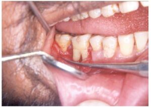
Once a horizontal component to the furca- tion has developed (class II), therapy becomes more complicated. Shallow horizontal involvement without significant vertical bone loss usually responds favorably to localized flap procedures with odontoplasty and osteo- plasty. Isolated deep class Il furcations may respond to flap procedures with osteoplasty and odontoplasty (Figure 68-6). This reduces the dome of the furcation and alters gingival contours to facilitate the patient’s plaque removal.
Classes II to IV: Advanced Defects.
The development of a significant horizontal component to one or more furcations of a multirooted tooth (late class IH, class III or IV) or the development of a deep vertical component to the furca poses additional problems. Nonsurgical treatment is usually ineffective because the ability to instrument the tooth surfaces adequately is compromised. Periodontal surgery, endodontic therapy, and restoration of the tooth may be required to retain the tooth.
SURGICAL THERAPY
Root Resection :-
Root resection may be indicated in multirooted teeth with grade II to IV furcation involvements. Root resection may be performed on vital teeth or endodontically treated teeth. It is preferable, however, to have endo- dontic therapy completed before resection of a root. If this is not possible, the pulp should be removed, the patency of the canals determined, and the pulp chamber medicated before resection. It is distressing for both patient and clinician to perform a vital root resection and subsequently have an untoward event occur, such as perforation, fracture of the root, or an inability to instrument the canal.
The indications and contraindications for root resection were well summarized by Bassaraba. In general, teeth planned for root resection include the following:
1. Teeth that are of critical importance to the overall dental treatment plan. Examples are teeth serving as abutments for fixed or removable restorations for which loss of the tooth would result in loss of the prosthesis and entail major prosthetic re-treatment.
2. Teeth that have sufficient attachment remaining for function. Molars with advanced bone loss in the interproximal and interradicular zones, unless the lesions have three bony walls, are not candidates for root amputation.
3. Teeth for which a more predictable or costeffective method of therapy is not available. Examples are teeth with furcation defects that have been treated successfully with endodontics but now present with a vertical root fracture, advanced bone loss, or caries on bone root.
4. Teeth in patients with good oral hygiene and low activity for caries are suitable for root resection. Patients unable or unwilling to perform good oral hygiene and preventive measures are not suitable candidates for root resection or hemisection. Root-resected teeth require endodontic treatment¹ and usually cast restorations.
These therapies can represent a sizable financial invest- ment by the patient in an effort to save the tooth. Alternative therapies and their impact on the overall treatment plan should always be considered and presented to the patient.
Which Root to Remove :-
A tooth with an isolated furcation defect in an otherwise intact dental segment may present few diagnostic problems. However, the existence of multiple furcation defects of varying severity combined with generalized advanced periodontitis can be a challenge to treatment planning. Careful diagnosis usually allows the therapist to determine the feasibility of root resection and the identification of which root to remove before surgery .
The following is a guide to determining which root should be removed in these cases:
1. Remove the root(s) that will eliminate the furcation and allow the production of a maintainable architecture on the remaining roots.
2. Remove the root with the greatest amount of bone and attachment loss. Sufficient periodontal attachment must remain after surgery for the tooth to withstand the functional demands placed on it. Teeth with uniform advanced horizontal bone loss are not suitable for root resection.
3. Remove the root that best contributes to the elimination of periodontal problems on adjacent teeth. For example, a maxillary first molar with a class III buccal-to-distal furcation is adjacent to a maxillary second molar with a two-walled intrabony defect between the molars and an early class II furcation on the mesial furcation of the second molar. There may or may not be local anatomic factors affecting the teeth. The removal of the distobuccal root of the first molar allows the elimination of the furcation and management of the two-walled intrabony lesion and also facilitates access for instrumentation and maintenance of the second molar .
4. Remove the root with the greatest number of anatomic problems, such as severe curvature, developmental grooves, root flutings, or accessory and multiple root canals.
5. Remove the root that least complicates future periodontal maintenance.
Hemisection :-
Hemisection is the splitting of a two-rooted tooth into two separate portions. This process has been called bicuspidization or separation because it changes the molar into two separate roots. Hemisection is most likely to be performed on mandibular molars with buccal and lingual class II or III furcation involvements. As with root resection, molars with advanced bone loss in the interproximal and interradicular zones are not good candidates for hemisection. After sectioning of the teeth, one or both roots can be retained. This decision is based on the extent and pattern of bony loss, root trunk and root length, ability to eliminate the osseous defect, and endodontic and restorative considerations. The anatomy of the mesial roots of mandibular molars often leads to their extraction and the retention of the distal root to facilitate both endodontic and restorative therapy.
The interradicular dimension between the two roots of a tooth to be hemisected is also important. Narrow interradicular zones can complicate the surgical proce- dure. The retention of both molar roots can complicate the restoration of the tooth, since it may be virtually impossible to finish margins or to provide an adequate embrasure between the two roots for effective oral hygiene and maintenance . Therefore, orthodontic separation of the roots is often required to allow restoration with adequate embrasure form. The result can be the need for multiple procedures and extensive interdisciplinary therapy. In such patients the availability of other treatment alternatives should be considered, such as guided tissue/guided bone regeneration or replacement by osseointegrated dental implants.
Root Resection/Hemisection Procedure :-
After appropriate local anesthesia, a full- thickness mucoperiosteal flap is elevated. Root resection or hemisection of teeth with advanced attachment loss usually requires opening both facial and lingual/palatal flaps. Typically, a root cannot be resected without elevating a flap. The flap should provide adequate access for visualization and instrumentation and minimize surgical trauma.
After debridement, resection of the root begins with the exposure of the furcation on the root to be removed . The removal of a small amount of facial or palatal bone may be required to provide access for elevation and facilitate root removal . A cut is then directed from just apical to the contact point of the tooth, through the tooth, and to the facial and distal orifices of the furcation . This cut is made with a high-speed, surgical-length fissure or cross-cut fissure carbide bur. The placement of a curved periodontal probe into or through the furcation aids in orienting the angle of the resection. For hemisection, a vertically oriented cut is made faciolingually through the buccal and lingual developmental grooves of the tooth, through the pulp chamber, and through the furcation. If the sectioning cut passes through a metallic restoration, the metallic portion of the cut should be made before flap elevation. This prevents contamination of the surgical field with metallic particles.
If a vital root resection is to be performed, a more horizontal cut through the root is advisable . An oblique cut exposes a large surface area of the radicular pulp and/or dental pulp chamber. This can lead to post- operative pain and can complicate endodontic therapy. A horizontal cut, although it may complicate root removal, has less postoperative complications. This root stump can be removed by odontoplasty after the completion of endodontic therapy or at the time of tooth preparation.
After sectioning, the root is elevated from its socket. Care should be taken not to traumatize bone on the remaining roots or to damage an adjacent tooth. Removal of the root provides visibility to the furcation aspects of the remaining roots and simplifies the debridement of the furcation with hand, rotary, or ultrasonic instruments. If necessary, odontoplasty is performed to remove portions of the developmental ridges and prepare a furcation that is free of any deformity that would enhance plaque retention or adversely affect plaque removal .
Patients with advanced periodontitis often have root resection performed in conjunction with other surgical procedures. provides an example of combining root resection and periodontal osseous surgery. The bony lesions that may be present on adjacent teeth are then treated using resective or regenerative therapies. After resection, the flaps are then approximated to cover any grafted tissues or slightly cover the bony margins around the tooth. Sutures are then placed to maintain the position of the flaps. The area may or may not be covered with a surgical dressing.
The removal of a root alters the distribution of occlusal forces on the remaining roots. Therefore it is wise to evaluate the occlusion of teeth from which roots have been resected and, if necessary, adjust the occlusion. Centric holds should be maintained, but eccentric forces should be eliminated from the area over the root that was removed. Patients with advanced attachment loss may benefit from temporary stabilization of the resected tooth to prevent movement .